Diagnosis and Associated Risks Factors

Acute RVO patients most commonly present with painless, blurred vision, vision loss or floaters.1 The initial exam should include visual acuity, pupillary assessment, slit-lamp exam looking for new iris vessels, and gonioscopy for evaluation of the iridocorneal angle.2 Dilated fundus exam may reveal intraretinal hemorrhages, dilated and tortuous retinal veins, retinal edema, exudates and cotton wool spots.3 As RVO patients may also have elevated ocular perfusion pressure or arteriovenous nicking with focal narrowing, optic nerve head (ONH) evaluation is necessary for all RVO patients to rule out glaucoma.3 Color and red-free fundus photography is useful to document the severity of the retina findings.4

Optical Coherence Tomography (OCT) is used to determine retinal thickness, monitor macular edema and vitreoretinal interface changes.2 Treatment decisions are commonly based on OCT measurements because they are objective and quantifiable.2 Visualization of microvasculature of the retina and choroid is possible utilizing Optical Coherence Tomography Angiography (OCTA), a non-invasive way to detect capillary nonperfusion, enlarged avascular zones and vascular abnormalities.2 Fluorescein angiography is useful in assessing retinal ischemia, delayed vein filling, the presence of retinal neovascularization with fluorescein leakage and collateral vessels.4

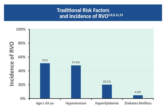
Clinicians need to educate their patients about risk factors that increase their chances of developing an RVO. Linkage of RVO to certain systemic vascular risks including hypertension, hyperlipidemia, diabetes, active smoking and peripheral vascular disease has been made.3,5-7 A meta-analysis found that 47.9% of RVO cases were attributed to hypertension, 20.1% to hyperlipidemia and 4.9% to diabetes.3,8 Hypertension can cause a thickening or hardening of the overlying artery which causes turbulent blood flow that is believed to promote venous thrombosis.9 The occlusion can obstruct blood flow and increase venous pressure.2,10 This may lead to vascular leakage and edema, hemorrhages and ischemia.9

Fifty-eight percent of patients with CRVO onset at an age younger than 50 were found to have a nontraditional risk factors, such as an underlying systemic disease, hypercoagulability or history of hormonal contraceptives on evaluation.11 In a cohort with systemic lupus erythematosus, the incidence of CRVO was 3.5 times higher than in a control population.10,11 Although men and women are affected equally, the incidence of RVO increases with age, with > 50 % of cases occurring in patients over 65 years old.9

Systemic factors that protect against BRVO include increased high density lipoprotein (HDL) levels, moderate alcohol consumption and increased exercise.3 In contrast, only increased physical activity has been shown to protect against CRVO.3,6 Communication with the primary care provider is essential, since patients with RVO may have a higher risk of cardiovascular disease and cerebrovascular accidents.3,12

RVO Risk Factor Assessments

References

  1. Schmidt-Erfurth U, Garcia-Arumi J, Gerendas BS, et al. Guidelines for the Management of Retinal Vein Occlusion by the European Society of Retinal Specialists (EURETINA). Ophthalmologica. 2019;242:123-162. https://www.karger.com/Article/Pdf/502041
  2. Flaxel CJ, Adelman RA, Bailey ST, et al. Retinal Vein Occlusions Preferred Practice Patterns. Ophthalmology. 2019;127:P288-P320. https://www.aaojournal.org/article/S0161-6420(19)32096-2/fulltext
  3. Jenkins T, Su D, Klufas MA. RVO Overview. Retina Today. April, 2018:40-58. https://retinatoday.com/articles/2018-apr/rvo-overview
  4. Scruggs B, Quist T, Kravchuk O, Sohn E. Branch Retinal Vein Occlusion, Eyerounds.org. Last updated July 7, 2018. https://webeye.ophth.uiowa.edu/eyeforum/cases/274-branch-retinal-vein-occlusion.htm
  5. Ehlers JP, Fekrat S. Retinal vein occlusion: Beyond the acute event. Surv Ophthalmol. 2011;56:281-299. https://pubmed.ncbi.nlm.nih.gov/21601903/
  6. Sperduto RD, Hiller R, Chew E, et al. Risk factors for hemiretinal vein occlusion: Comparison with risk factors for central and branch retinal vein occlusion: The Eye Disease Case-Control study. Ophthalmology. 1998;105:765-771. https://pubmed.ncbi.nlm.nih.gov/9593373/
  7. Bertelsen M, Linneberg A, Christoffersen N, Vorum H, Gade E, Larsen M. Mortality in patients with central retinal vein occlusion. Ophthalmology. 2014;121:637-642. https://pubmed.ncbi.nlm.nih.gov/24053999/
  8. O’Mahoney PRA, Wong DT, Ray JG. Retinal vein occlusion and traditional risk factors for atherosclerosis. Arch Ophthalmol. 2008;126:692-699. https://pubmed.ncbi.nlm.nih.gov/18474782/
  9. Morris R, Retinal vein occlusion, Kerala J Ophthalmol. 2016;28:4-13. https://www.kjophthal.com/text.asp?2016/28/1/4/193868
  10. Wills Eye Hospital. Central Retinal Vein Occlusion (CRVO). https://www.willseye.org/central-retinal-vein-occlusion-crvo/ Accessed 12/14/20.
  11. Rothman A, Thomas A, Khan K. Central Retinal Vein occlusion in young individuals: A comparison of risk factors and clinical outcomes. Retina. 2019;39:1917-1924. https://pubmed.ncbi.nlm.nih.gov/30085977/
  12. Chen YY, Sheu SJ, Hu HY, Chu D, Chou P. Association between retinal vein occlusion and an increased risk of acute myocardial infarction: A nationwide population-based follow-up study. PloS One. 2017;12:e0184016 https://pubmed.ncbi.nlm.nih.gov/28898259/
  13. Jumper JM, RVO workup: When it’s necessary and what to order. Retinal Specialist. March 15, 2015. https://www.retina-specialist.com/article/rvo-workup-when-its-necessary-and-what-to-order

Pin It on Pinterest

Directory
Scroll to Top

Scientific Council

Neil M. Bressler, MD

James P. Gills Professor of Ophthalmology
Professor of Ophthalmology, Johns Hopkins University School of Medicine
Wilmer Eye Institute, Johns Hopkins Medicine
Baltimore, MD

A. Paul Chous, MA, OD, FAAO

Specializing in Diabetes Eye Care & Education, Chous Eye Care Associates
Adjunct Professor of Optometry, Western University of Health Sciences
AOA Representative, National Diabetes Education Program
Tacoma, WA

Steven Ferrucci, OD, FAAO

Chief of Optometry, Sepulveda VA Medical Center
Professor, Southern California College of Optometry at Marshall B. Ketchum University
Sepulveda, CA

Julia A. Haller, MD

Ophthalmologist-in-Chief
Wills Eye Hospital
Philadelphia, PA

Allen C. Ho, MD, FACS

Director, Retina Research
Wills Eye Hospital
Professor and Chair of the Department of Ophthalmology
Thomas Jefferson University Hospitals
Philadelphia, PA

Charles C. Wykoff, MD, PhD

Director of Research, Retina Consultants of Houston
Associate Professor of Clinical Ophthalmology
Blanton Eye Institute & Houston Methodist Hospital
Houston, TX

Clinician Scientific & Educational Resources

The RELIEF Clinical Toolkit is an online tool that aims to provide clinicians with up-to-date information on the presentation, prognosis, pathophysiology, and treatment strategies for Retinal Vein Occlusion (RVO) in patients with diabetes who have or are at risk for developing RVO. Click on one of the options below to learn more about RVO.

This activity is provided by Med Learning Group.
This activity is supported by an educational grant from Regeneron Pharmaceuticals, Inc.

Copyright © 2019 | Retinal Vein Occlusion | All Rights Reserved | Website by Divigner