RVO Management

Retinal vein occlusion (RVO) management for patients involves treating any complications due to the vein occlusion in addition to systemic disease etiology, such as hypertension, hyperlipidemia and diabetes (known risk factors for RVO).1-3 A meta-analysis and systematic review suggests patients with any RVO have an increased risk of cardiovascular events and all-cause mortality.4,5 Therefore, as part of retinal vein occlusion management, the eye physician should refer patients with a RVO to their primary care provider (PCP) for appropriate management of their systemic condition(s). The eye physician should also communicate all eye examination results.4,6

Central retinal vein occlusion (CRVO)

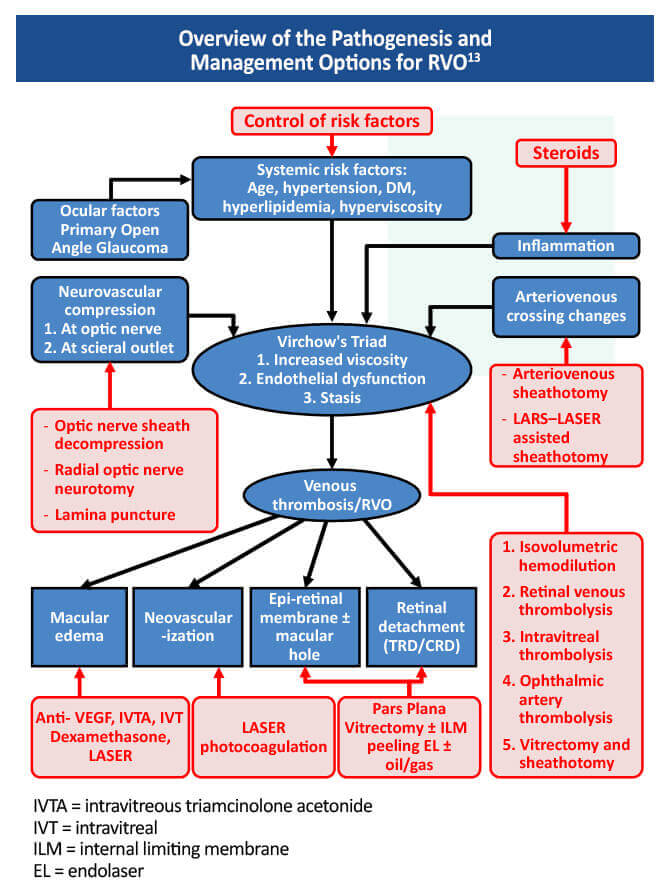
The ischemic subtype of CRVO accounts for about 20% of cases and is associated with worse initial presenting visual acuity (VA) and poor visual prognosis even after edema resolution.7,8 The irreversible loss of VA in macula edema is usually attributed to permanent loss of photoceptor cells.9 In 2019, the AAO recommended intravitreal anti-VEGF vision loss therapy as first-line treatment for macular edema secondary to RVO, reserving corticosteroids as second-line treatment due to secondary glaucoma and cataract formation. Panretinal laser photocoagulation has also been recommended in the setting of neovascular complications secondary to RVO.4

A common first-line vision loss treatment protocol for CRVO patients would include a loading dose of monthly injections over 3 months of an vascular endothelial growth factor (VEGF) inhibitor, such as ranibizumab or aflibercept (or off-label bevacizumab).8 Monthly treatment should continue until visual and anatomical outcomes are stable over 3 consecutive monthly assessments.10 For individuals who develop iris neovascularization or retinal neovascularization following development of CRVO, the best treatment is dense peripheral panretinal photocoagulation (PRP).4,11

Branch retinal vein occlusion (BRVO)

Branch retinal vein occlusion management (BRVO) depends on visual acuity, macular edema and ischemic changes.1,12 Patients with a non-ischemic BRVO and VA of 20/40 or better should be monitored in 3-month intervals.10 Monthly anti-VEGF injections should be initiated if VA is worse than 20/40 or if there is macular edema.10

Retinal Vein Occlusion Management

References

  1. Jenkins T, Su D, Klufas M. RVO Overview. Retina Today. April, 2018:40-58. https://retinatoday.com/articles/2018-apr/rvo-overview
  2. O’Mahoney PRA, Wong DT, Ray JG. Retinal vein occlusion and traditional risk factors for atherosclerosis. Arch Ophthalmol. 2008;126:692-699. https://pubmed.ncbi.nlm.nih.gov/18474782/
  3. Flaxel CJ, Adelman RA, Bailey ST, et al. Retinal Vein Occlusions Preferred Practice Patterns. Ophthalmology. 2019;127:P288-P320. https://pubmed.ncbi.nlm.nih.gov/31757503/
  4. Wills Eye Hospital. Central Retinal Vein Occlusion (CRVO). https://www.willseye.org/central-retinal-vein-occlusion-crvo/
  5. Guyatt GH, Oxman AD, Vist GE, et al. GRADE: An emerging consensus on rating quality of evidence and strength of recommendations. BMJ. 2008;336:924-926. https://pubmed.ncbi.nlm.nih.gov/18436948/
  6. O’Mahoney PR, Wong DT, Ray JG. Retinal vein occlusion and traditional risk factors for atherosclerosis. Arch Ophthalmol. 2008;126:692-699. https://pubmed.ncbi.nlm.nih.gov/18474782/
  7. Hayreh SS, Podhajsky PA, Zimmerman MB. Natural history of visual outcome in central retinal vein occlusion. Ophthalmology. 2011;118:119-133.e2. https://pubmed.ncbi.nlm.nih.gov/20723991/
  8. Ashraf M, Souka A, Singh R. Central retinal vein occlusion: Modifying current treatment protocols. Eye (Lond). 2016;30:505-514. https://pubmed.ncbi.nlm.nih.gov/26869163/
  9. Lardenoye C, Probst K, Jaap K. Photoceptor function in eyes with macular edema. Invest Opthalmol Vis Sci. 2000,41:4048-4053. https://pubmed.ncbi.nlm.nih.gov/11053311/
  10. Morris R, Retinal Vein Occlusion. Kerala J Ophthalmol. 2016;28:4-13. https://www.kjophthal.com/text.asp?2016/28/1/4/193868
  11. A randomized clinical trial of early panretinal photocoagulation for ischemic central vein occlusion. The Central Vein Occlusion Study Group N report. Ophthalmology. 1995;102:1434-1444. https://pubmed.ncbi.nlm.nih.gov/9097789/
  12. Stuart A. Untangling retinal vein occlusion. Eyenet Magazine. November, 2013. https://www.aao.org/eyenet/article/untangling-retinal-vein-occlusion
  13. Mishra D, Ramanjulu R, Shanmugam M. Retinal vein occlusion. eOphtha.com. Accessed December 20, 2020. http://www.eophtha.com/posts/retinal-vein-occlusion
 

Pin It on Pinterest

Directory
Scroll to Top

Scientific Council

Neil M. Bressler, MD

James P. Gills Professor of Ophthalmology
Professor of Ophthalmology, Johns Hopkins University School of Medicine
Wilmer Eye Institute, Johns Hopkins Medicine
Baltimore, MD

A. Paul Chous, MA, OD, FAAO

Specializing in Diabetes Eye Care & Education, Chous Eye Care Associates
Adjunct Professor of Optometry, Western University of Health Sciences
AOA Representative, National Diabetes Education Program
Tacoma, WA

Steven Ferrucci, OD, FAAO

Chief of Optometry, Sepulveda VA Medical Center
Professor, Southern California College of Optometry at Marshall B. Ketchum University
Sepulveda, CA

Julia A. Haller, MD

Ophthalmologist-in-Chief
Wills Eye Hospital
Philadelphia, PA

Allen C. Ho, MD, FACS

Director, Retina Research
Wills Eye Hospital
Professor and Chair of the Department of Ophthalmology
Thomas Jefferson University Hospitals
Philadelphia, PA

Charles C. Wykoff, MD, PhD

Director of Research, Retina Consultants of Houston
Associate Professor of Clinical Ophthalmology
Blanton Eye Institute & Houston Methodist Hospital
Houston, TX

Clinician Scientific & Educational Resources

The RELIEF Clinical Toolkit is an online tool that aims to provide clinicians with up-to-date information on the presentation, prognosis, pathophysiology, and treatment strategies for Retinal Vein Occlusion (RVO) in patients with diabetes who have or are at risk for developing RVO. Click on one of the options below to learn more about RVO.

This activity is provided by Med Learning Group.
This activity is supported by an educational grant from Regeneron Pharmaceuticals, Inc.

Copyright © 2019 | Retinal Vein Occlusion | All Rights Reserved | Website by Divigner